Weiwei Xia* Xianghua Zeng*,
College of Physics Science and Technology & Institute of Optoelectronic Technology ,Yangzhou University,
Yangzhou 225002, P.R. China
Vertically aligned quasi two-dimensional (2D) ZnO nanorods (NRs) on carbon fiber paper were prepared by a modified hydrothermal approach, where the ZnO nanorods with a diameter of 100 to 200 nm and length of ~ 1um are uniformly distributed on the surface of the carbon paper. To design visible light detector, intact ZnO@ZnS cores-hell hierarchal structures with an outer ZnS shell of ~25 nm were fabricated under an elaborate sulfidation treatment. Photoluminescence spectra exhibited that the bare ZnO nanorods have a sharp band emission at 380 nm and a strong broad mission around 580 nm, whereas the near band emission at 380 nm was depressed and a visible light emission around 500 nm was greatly enhanced after a sulfidation treatment. XPS and ESR measurements were carried out to examine the constituents and the defects of the prepared samples. Self-powered photoelectrochemical cell-type detector was designed by using ZnO and ZnO@ZnS NRs as anode electrodes under an illumination of 564 ± 60 nm, the photocurrent of ZnO@ZnS NRs was obtained as 6.79 µA with an on-off interval of 40 s at a bias of 0 V for 60 mW/cm2, which is ten times larger than the current 0.67µA of the bare ZnO electrode. The enhanced visible-light photoresponse performance could be attributed to the hierarchal structures and the good conductivity of the carbon fiber paper. As the quasi 2D core-shell hierarchal structures will provide both more efficient active sites as it exposed more catalytic sites to the electrolyte, and more charge carriers from the reduced work function at the interface between two semiconductors.
Keywords: ZnO/ZnS core-shell, carbon substrate, visible light, self-powered photodetector
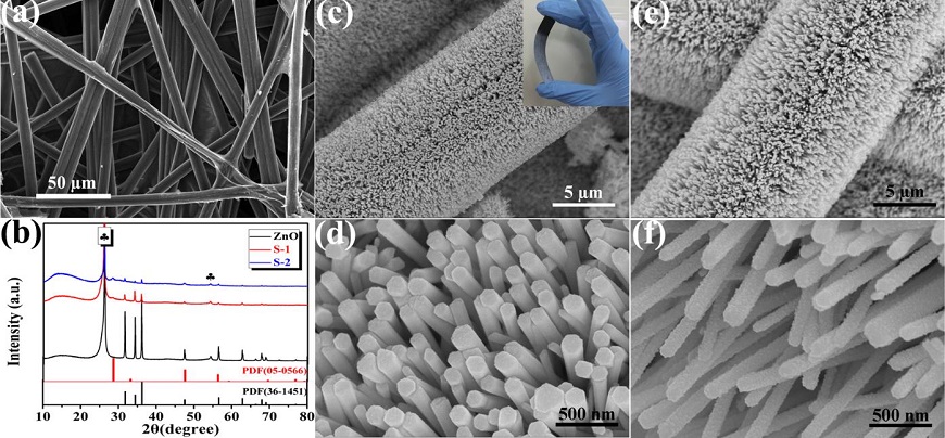
Figure 1 (a) SEM image of bare carbon fiber paper; (b) XRD patterns of the as-obtained samples; (c) and (d) are low and high magnification SEM images of the ZnO nanorod array grown on carbon fiber paper; (e) and (f) are low and high magnification SEM images of sample S-2.
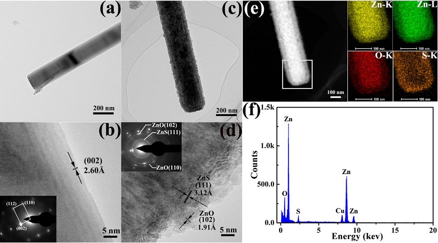
Figure 2 (a-b) Low and higher magnification TEM images with inset for corresponding SAED pattern of as-selected single ZnO nanorod. (c-d) Typical TEM images of a single ZnO@ZnS core-shell nanorod with different magnification. (e) TEM image of an individual ZnO-ZnS nanorod with EDS elemental distribution mapping of Zn, O, and S respectively. (f) the corresponding EDX spectrum of the product.

Figure 3 (a) The schematic illustration of self-powered PEC type detector; (b) and (c) Photocurrent responses under on/off of 40 s at 0.0 V vs. Ag/AgCl with illumination of 564 ± 60 nm 60 mW/cm2 incident intensity; (d) Transient decay times of two ZnO@ZnS samples measured at the same condition.

